You have strange pains in your abdomen. You suspect your child has broken a bone. You were in a car accident and your doctor is concerned about internal injuries. You’re expecting a baby.
In every instance, you’ll need medical imaging, which can help to reveal what’s happening inside your body. There are several kinds of imaging, each using different machines and appropriate for different purposes, and it’s helpful to know the test you’ll receive so you know how to prepare and what to expect.
UNC Health pediatric radiologist Carolina Guimaraes, MD, explains the types of imaging tests your doctor might order.
X-ray (Radiograph)
If you’re in the emergency department or urgent care, it’s common to have an X-ray, or radiograph, as the first step for imaging.
“Radiographs are inexpensive and fast, great for the evaluation of bone fractures and acute lung abnormalities such as pneumonia,” Dr. Guimaraes says.
X-rays send a very small amount of ionizing radiation through your body; your bones, organs and tissue absorb the radiation in different ways, which is shown in the image created by the machine. Because of the way bones absorb radiation, they look very bright in the image, making it easy to see how and where a bone is affected.
Organs such as lungs appear gray; an X-ray can help to identify the presence of an infection or other abnormality. If you have pneumonia, for example, a chest X-ray can show the presence of fluid or pus in the lung, or if your child swallowed a metallic object, it will show up on an image of their abdomen.
Any part of the body can be X-rayed, and this test doesn’t require any special preparation. No equipment touches the patient.
There are limits to the level of detail that an X-ray can show, so Dr. Guimaraes says you may need a more in-depth imaging study if an X-ray doesn’t reveal the source of your symptoms.
Ultrasound
An ultrasound uses sound waves, not radiation, to create real-time images of the inside of the body. A technologist will apply gel and use an ultrasound wand to examine the relevant area.
Ultrasounds can help your doctor to visualize some parts of the body that don’t show up as well on an X-ray.
“It’s often used for visualization of abdominal organs or superficial lumps,” Dr. Guimaraes says.
Ultrasounds can be used to diagnose issues with the abdominal organs (stomach area), pelvic organs, breasts, thyroid and prostate. They can see veins and evaluate blood flow. It’s also how doctors monitor the growth and development of your baby during pregnancy.
As with X-ray, there may be some limits as to what can be visualized with an ultrasound. Sound waves don’t travel well through bones or gas, which can affect what’s seen, so you might also require another test.
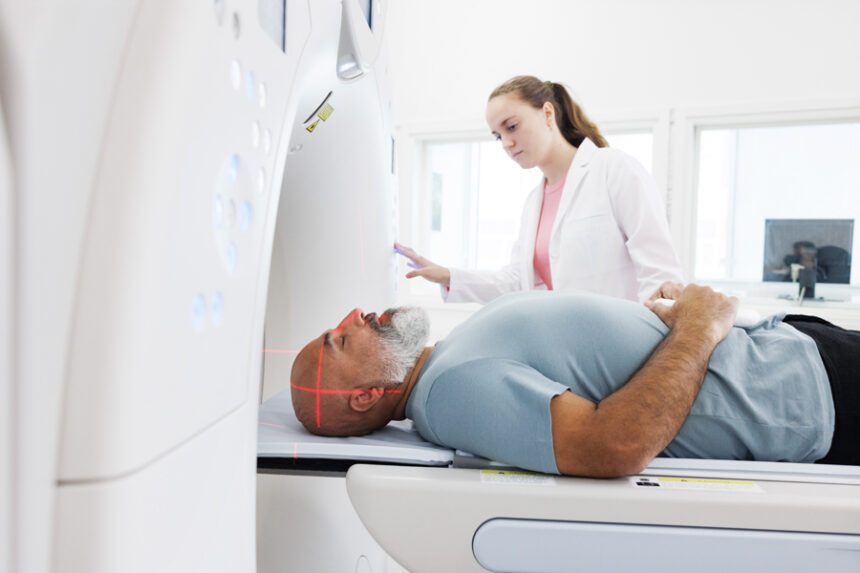
Computed Tomography (CT)
Computed tomography, also referred to as a CT or CAT scan, uses ionizing radiation, as X-rays do, but with a different type of scanner and a computer that creates more precise images. It can be used for a variety of concerns, including bones, organs, vessels and soft tissues.
“With CT, you can get more detail of internal structure, a level of detail that X-ray doesn’t give you,” Dr. Guimaraes says. “It’s also a very fast study, commonly used in trauma cases. You can image a patient’s entire body in a couple of minutes and plan for important next steps such as surgery, if needed.”
During a CT scan, you lie on a table and are moved through a doughnut-shaped scanner; because of that shape, CT produces three-dimensional images and cross-sections of what’s inside the body, as opposed to an X-ray, which is like taking a two-dimensional photograph. The scanner is open, not a long tube, meaning the risk of claustrophobia is low as your entire body isn’t inside it at once.
Dr. Guimaraes says for some CT scans, you might need to drink contrast or have an IV placed so that contrast can be administered. Contrast is a dye that helps to improve the visibility of parts of your body, which helps to create a better image.
CT does use more ionizing radiation than X-ray; for example, a chest X-ray exposes you to a dose of 0.1 millisieverts (mSv) while a chest CT requires 6.1 mSv. The average person is exposed to 3 mSv per year by simply going about daily life; a flight from the east coast to the west coast of the United States exposes you to 0.035 mSv. Because large cumulative doses of this type of radiation have been linked to increased incidence of cancer, Dr. Guimaraes says that providers are mindful about when they use CT.
“We always weigh benefits and risks,” she says. “We don’t do studies that aren’t necessary. We try to start with X-ray or ultrasound. If the test is important for the patient, we do it at the lowest dose of radiation possible for the best diagnosis.”
Dr. Guimaraes says technology continues to improve and that the doses of radiation needed are becoming lower, minimizing long-term risk.
Magnetic Resonance Imaging (MRI)
An MRI doesn’t use radiation; instead, it uses magnetic fields and radio waves to create high-resolution images.
“An MRI is our most detailed study because it can show the best differentiation between body tissue,” Dr. Guimaraes says.
Though it shows the most detail, it’s likely not going to be your doctor’s first choice because it is more costly than other tests, and it takes more time. While other tests take a few minutes, most MRIs usually take 30 minutes to an hour, which may not be appropriate following a trauma or when you know a less expensive test will provide the necessary information.
MRIs can also be difficult for some people.
“You have to stay really still the entire time or the images are not adequate,” Dr. Guimaraes says. “Some people can feel claustrophobic, because it’s a narrower, longer tube than CT. Especially when imaging the brain, the machine can feel close to your head and it is also very noisy.”
Young children may need sedation if they need an MRI as they may not be able to keep still; if you’re an adult and worried about claustrophobia, talk to your doctor. They may prescribe a medication for anxiety. You’ll be given ear plugs for the noise, and you may be able to listen to music. Children may also watch a movie.
As with CT, you may need an IV with contrast placed before your imaging.
Fluoroscopy
If an X-ray is like taking a photo of your insides, then a fluoroscopy is like watching a video of them.
“Fluoroscopy gives us real-time continuous X-rays, rather than one static image,” Dr. Guimaraes says, which means that your doctor can see whether your body’s internal systems are functioning correctly by watching them in action. This can be used to diagnose swallowing problems, gastrointestinal issues, urinary tract abnormalities, or to guide placement of catheters, stents or other devices.
“A common scenario is that you’d take contrast, stand or lay near an X-ray tube, and we can watch the contrast make its way through the digestive system,” Dr. Guimaraes says.
Mammogram
A mammogram uses a low dose of radiation (a similar amount to an X-ray) to create an image of the internal breast tissue, which can allow your doctor to detect breast cancer. During the mammogram, a trained technologist compresses the breasts briefly by the scanner to take the best image.
The American College of Radiology (ACR) recommends that women get a screening mammogram starting at age 40. Breast cancer is very common, and most women do not have a family history of breast cancer. Knowing your personal risk for developing breast cancer is very important; the ACR recommends you have a risk assessment by the age of 25 to decide if you may need to start screening earlier or with other tests, such as MRI.
Almost half of women will have dense breast tissue on their mammogram, which will be noted in your results. If you have dense tissue, you may also benefit from other testing.
You may need diagnostic imaging if you feel a lump, have skin changes or nipple discharge, or had an abnormal screening mammogram. Diagnostic imaging may be a mammogram and/or a breast ultrasound, which take longer as the technologist will create more detailed images of the area of concern.
Nuclear Medicine
Nuclear imaging involves injecting a small amount of radioactive material into your veins that will spread through your bloodstream to create images of internal organs. This uses a special gamma camera, sometimes in combination with MRI or CT. The camera may stay still on top of you or rotate around your body, but it does not touch you. Pictures may be taken on the same day of the radiotracer injection or days after.
“This scan is most commonly used for cancer detection and monitoring,” Dr. Guimaraes says; it can also be used for patients with heart disease and some brain disorders.
If there are diseased cells, they will absorb a greater amount of the radioactive material. That means these images show your doctor how a particular organ might be functioning, rather than just providing an image of the structure and any abnormalities.
No matter what type of test you’re having, Dr. Guimaraes encourages you to ask your provider questions if you have them. Make sure you understand the pre-scan instructions, such as whether you need to take off your jewelry, abstain from food or drink, or avoid wearing deodorant or lotion.
“Radiology nurses, technologists or doctors can explain the exam prior to imaging or do a simulation if you’re nervous,” she says, noting that child life specialists can help with children’s tests. “They can also explain why other studies are needed if your first one doesn’t give us the answers we need. Sometimes others are needed, but the different modalities usually complement each other.”
If you have questions about any type of medical test, talk to your doctor. If you need a doctor, find one near you.